|
|
(a)
|
|
(b)
|
(a): Quadrilateral surface meshes of the nerve (flattish top) and muscular (folds)
cell membranes at a neuromuscular junction.
The interior is also meshed into hexahedral elements.
(b): the mesh in (a) after quality improvement using geometric flow. The closeups
between (a) and (b) highlight the improvement in the shape of the quadrilaterals on
the membrane surface.
|

|

|

|

|

|
|
(c)
|
(d)
|
(e)
|
(f)
|
| |
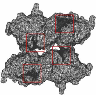
A Gaussian blurred volume map of an atomic resolution molecular model of mouse
acetylcholinesterase (mAChE). The region near the active site of mAChE (narrow gorge)
is highlighted by red box in all the figures. (a): output adaptive triangle mesh and
a closeup of the mesh near the gorge. (b): the same mesh, in a closeup,
after performing quality improvement.
(c,d): cross section of the interior and exterior hexahedral mesh of the same 3D map of mAChE.
Note the mesh adaptivity near the gorge.
(e,f): triangulated wireframe and tetrahedral mesh of a tetrameric cluster of mAChE (mAChE4).
The molecule predominantly occurs in such tetrameric forms, especially in
the synaptic cleft of neuromuscular junctions.
 click here for an interactive view of mAChE Model (viewer works with I.E only)
Additional
information for Neuro-Muscular Junction(NMJ) project
|
|
|
|
|