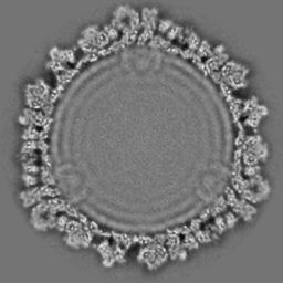
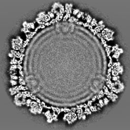
The images obtained from the cryo-electron microscopy are usually very noisy and have
very low contrast. It is quite necessary to smooth the noise as well as enhance the contrast
before other tasks can be conducted. This also happens right after the 3D electron density
maps are reconstructed. We have developed an adaptive contrast enhancement technique
and a PDE-based anisotropic diffusion technique for noise removal. The following pictures
show an example of our approaches on the reconstructed electron density map of Rice
Dwarf Virus (RDV). More details are available later or upon request. The Cryo-EM
images or the reconstructed maps may also be improved by Contrast Transfer
Function (CTF) correction. However, we are not working on this.
 |
 |
 |
| Fig. 1 Left: original image |
Middle: after anisotropic filtering |
Right: after contrast enhancement |
 |
 |
| Fig. 2 Left: original image |
Right: after anisotropic filtering |
|