| Biography | Research | Publications | Teaching | Group | Projects | Software | Sponsors | Collaborators |

|

|
|

|
| (a) | (b) | (c) | (d) |

|

|
|
|
| (e) | (f) | (g) | (h) |
|
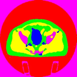
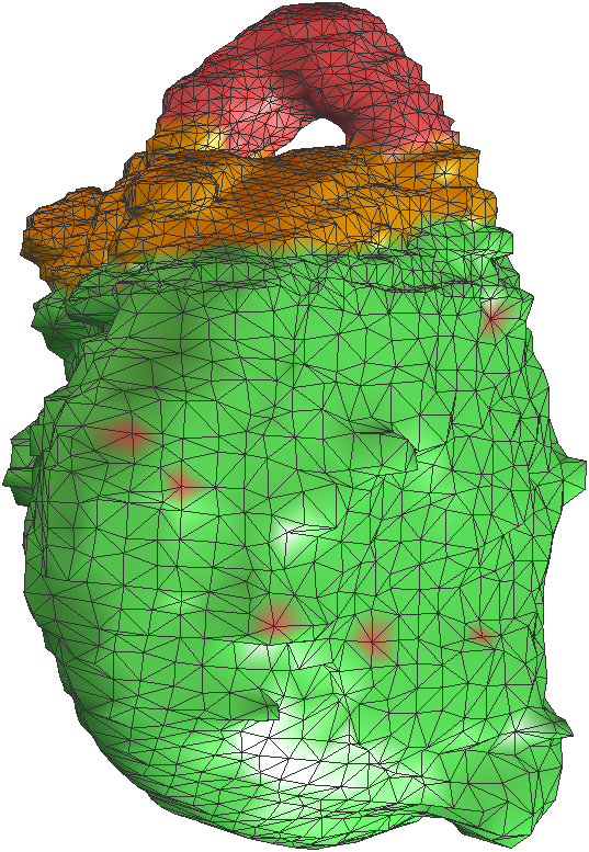
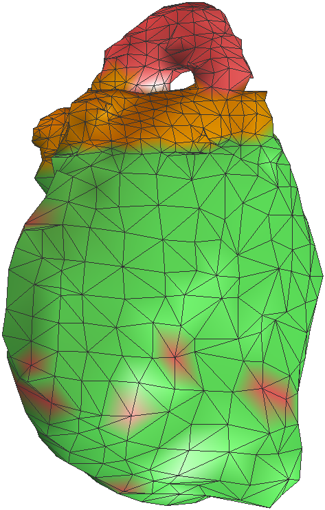
Abdominal CT scan is processed via our meshing pipeline. The focus of study in this image is the bladder and its volume occupancy and absorption of radiation, when in line with radiation beams to the prostate. (a): a slice of the original CT scan is shown. (b): same slice after our filtering step. (c): result of classification of the anatomic subregions, automatically segmented and colored differently (cross-section slice). (d): volume rendering of the abdominal region of the classified 3D image of the bladder, again shown in blue. (e): the bladder meshed using our hex mesher. Note the non-smoothness which leads to poor quality quadrilateral elements. (f): same mesh after quality improvement using geometric flow techniques. (g): the adaptive triangle mesh of the bladder. The hook (red) has been artificially chosen to require finer mesh adaptivity. (h): the same mesh as (g) after coarsening. Note the adaptive mesh density after coarsening. Additional images for Radiation Therapy Bio-Med studies computational modeling, simulation and visualization of anatomical and physiological processes. Here are Bio-Med projects information and gallery |
|||