| Biography | Research | Publications | Teaching | Group | Projects | Software | Sponsors | Collaborators |
|
|
|

|
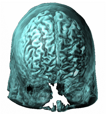
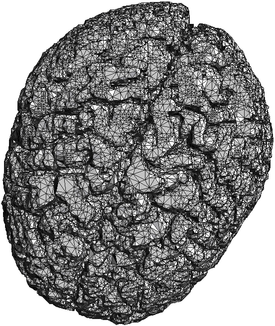
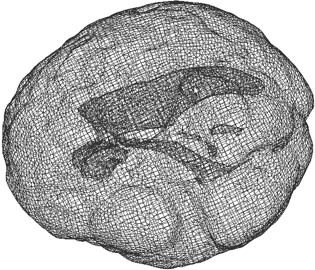
| (a) | (b) | (c) | (d) |
|
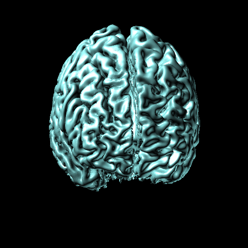
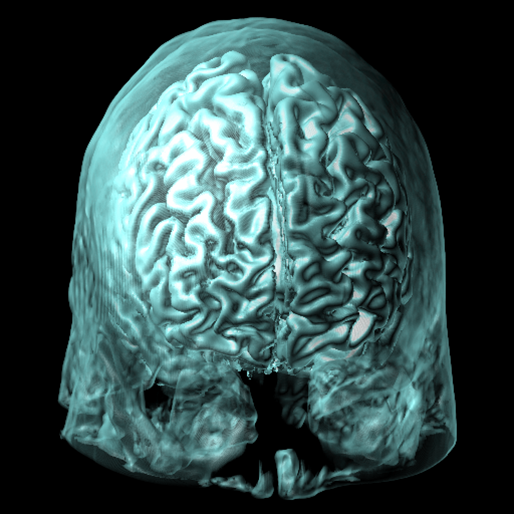
(a): MRI of a human head -the 3D image has been classified and a proper opacity function is automatically generated to distinguish the brain cortex in this volume rendered image. (b): the brain is segmented from the MRI, and a tetrahedral mesh is generated of the brain cortex and interior (including brain ventricle). (c): quadrilateral wireframe rendering showing the mesh interior of a simplified version of the brain mesh. Note the mesh adaptivity near the inner ventricles. (d): surface quadrilateral mesh (16874 quads) of the simplified version of the brain cortex being displayed. |
|||
|
|
|
|
| (e) | (f) | (g) | (h) |
|
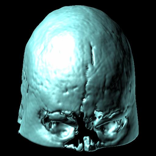
the fast informative visulaization of volumetric imaging data applying multi-transfer function. (e) shows outer surface of brain. (f) shows inside of brain. (g) shows both surface and inside. (h) shows a smooth surface from a surface triangulation by implicit triangular surface patches |
|||
|
|
|
| (i) | (j) | (k) |
|
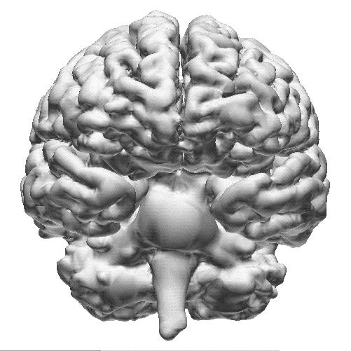
Brain Segmentaion with (i) front view, (j) top view, and (k) side view. |
||
|
|